REGISTER
LOG IN
HOME
RESOURCES
Publications
Conference Calendar
Past Quiz Results
ARTICLES
Revisiting ISHAM Asia 2021
The first AFWG CaseClinic is now live!
First-ever Study of Mycology Lab Practices in Asia
New Diagnostic Mycology E-learning Course
Antifungal prophylaxis: Whom, what and when
Fereydounia khargensis: A New Opportunistic Yeast Reported from Malaysia
9 Years of MMTN: Improving Fungal Disease Management in Asia Pacific
Echinocandins: Clinicians' Guide
Five controversies in mycology
Fungemia blood culture media
Deep dermatophytosis
AFWG Education Module 4: Is Antifungal Susceptibility Testing Useful for Clinical Management?
AFWG Education Module 5: TDM of Antifungal Agents - Essential or Optional?
AFWG Education Module 6: Antifungal Stewardship
10 common mistakes in laboratory mycology
Itraconazole: A Quick Guide for Clinicians
Evolving Fungal Landscape in Asia
10 common mistakes in clinical mycology
Laboratory Diagnosis of Pythiosis
ICMR Issues C. auris Advisory
Strengths and Limitations of Imaging for Diagnosis of IFI
Candidemia: Lessons Learned from Asian Studies for Intervention
Spotting invasive pulmonary aspergillosis in COVID-19 patients
Pivotal Asian Invasive Mold Study
Impact of the COVID-19 pandemic on IFI epidemiology and trends
Mycetoma in Asia: Still veiled in mystery
Cryptococcosis
Identifying IFI risk factors in patients with COVID-19
ASID ANZMIG x AFWG: Fungal Frontiers in the Asia Pacific – Webinar 2
New Antifungal Agents
Gilead IFI Masterclass: Current updates on the management of IFIs in immunocompromised hosts
The AFWG Masterclass: Advanced fungal education at your fingertips
A challenging case: A crisis unfolds
The role of antifungal stewardship in improving IFI outcomes
Making Precise Diagnoses: Experience from the Laboratory Skills Enhancement Course
A challenging case: A 68-year-old man with nasal and palatal ulcers
AFWG Online Education Module 3: Optimizing Dosing in IFI Management
AFWG Online Education Module 2: Antifungal Prophylaxis in Solid Organ Transplantation
AFWG Education Module 1: The Value of Clinical Mycology Laboratories
How do I interpret Candida in the urine?
How do I interpret Candida in respiratory tract cultures?
Cryptococcosis in HIV and non-HIV infected patients
Human Pythiosis
AFWGOnline Privacy Policy has been Updated
Management of fungal infections in high-risk patients
Striving for Perfection: Experience from the Laboratory Foundation Training Course
Know your fungal landscape in Vietnam
Recent Advances of Fungal Diagnostics in Asian Laboratories
Deep Dermatophytosis: A Case Report
Management of cryptococcosis and talaromycosis
A challenging case: A 49-year-old woman with sarcoidosis
Emerging yeast infections in Asia
Outbreak of Superbug Candida auris: Asian Scenario and Interventions
Championing Medical Mycology: Thoughts on the AFWG Laboratory Skills Enhancement Course
Mucormycosis and Pythiosis – New Insights
AML and the high risk of multiple infectious complications
Do We Need Modification of Recent IDSA & ECIL Guidelines while Managing Patients in Asia?
A hospital’s experience with candidemia and empirical therapy
Top 5 most viewed AFWG videos on YouTube
Fungal Academy 2015
Fluconazole in 2015
Fungal isolation protocol
Influencing Aspergillus
Fungal Asthma
Aspergillus
Laboratory Diagnosis of IPA
Two-Hot-to-Handle
Voriconazole
Educational Organizations
Literature Updates
 

Articles

A Challenging Case: A 68-year-old Man With Nasal And Palatal Ulcers

Share this
page


Share
this page

Dr Atul Patel, MD, FIDSA
Chief Consultant and Director
Infectious Diseases Clinic
Vedanta Institute of Medical Sciences
Ahmedabad, India

Clinical presentation

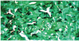
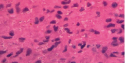
A 68-year-old patient with diabetes and chronic kidney disease was presented with fever, progressive nasal ulcer with crusting and painful palatal ulcer (Figure 1A). His work up for fever showed bilateral enlarged adrenal glands on abdominal CT scan. Biopsy from the palatal ulcer showed intracellular yeasts consistent with a diagnosis of histoplasmosis (Figures 1B & 1C). As Histoplasma antigen test was not available, a serum galactomannan test was conducted and yielded 3.48 ng/mL.

Figure 1. Clinical presentation of the patient with progressive nasal ulcer with crusting and painful palatal ulcer (A); and silver stain (B) and hematoxylin and eosin (H&E) stain (C) of the biopsy sample from the palatal ulcer

A
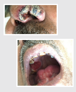
Clinical presentation of the patient with progressive nasal ulcer with crusting and painful palatal ulcer

B

Silver Stain

C

hematoxylin and eosin (H&E) stain

The patient was treated with oral itraconazole and had a good clinical response with marked regression in nasal and palatal ulcers (Figure 2) and a decrease in serum galactomannan level (2.48 ng/mL) after 6 weeks of therapy. The patient had complete clinical response after 7 months on antifungal treatment with further decrease of the serum galactomannan level to 1.45 ng/mL. His antifungal treatment was stopped at 9 months when the serum galactomannan level was at 0.68ng/mL. The patient had no sign of relapse 3 months follow-up after treatment completion. This case showed positive correlation of serum galactomannan levels with clinical response for histoplasmosis.

Figure 2. Marked regression in the nasal and palatal ulcers after 6 weeks on oral itraconazole therapy

Marked regression in the nasal and palatal ulcers after 6 weeks on oral itraconazole therapy

Discussion

The diagnosis of histoplasmosis is challenging as serological tests are not reliable, and culture is positive in only 50–70% and takes several weeks before it becomes positive.1

Detection of Histoplasma antigen from serum, urine and CSF is a sensitive and specific assay for diagnosing histoplasmosis but it is not available in many parts of world, including India. It may also be negative in localized diseases and only positive in progressive disseminated form of histoplasmosis. Histoplasmosis shares many clinical presentations and radiological features of tuberculosis, eg, fever of unknown origin, lymphadenopathy, adrenal gland enlargement, pulmonary nodules, etc. In regions of high tuberculosis prevalence, like India, histoplasmosis is generally not suspected. Diagnosis of histoplasmosis is generally made from the demonstration of intracellular yeasts in histopathological examination.

Positive Aspergillus galactomannan test can be used as a surrogate marker of Histoplasma infection. The minimum cutoff threshold for serum galactomannan to detect histoplasmosis is 0.4, giving a sensitivity of 82.1% and specificity of 100%.2 False-positive results from Aspergillus galactomannan assay has been reported in solid organ transplant recipients with histoplasmosis.3 Cross-reactivity between Histoplasma antigen and Aspergillus galactomannan has also been described in an HIV-infected patient with histoplasmosis.4 Therefore, physicians need to differentiate the 2 disease entities, especially when it comes to treatment decision making. In countries where Histoplasma antigen assay is not available, the cross-reactivity between Histoplasma and galactomannan antigens could be helpful surrogate marker for making a diagnosis of histoplasmosis in appropriate clinical and laboratory settings.

Clinical pearls

  • Aspergillus galactomannan assay with a cutoff ODI of 0.4 can be used as a reliable surrogate marker to detect for histoplasmosis
  • Decline in serum galactomannan correlates with clinical response in patient with histoplasmosis
  • Cross-reactivity between Aspergillus galactomannan and Histoplasma antigen can lead to false-positive results especially in immunocompromised patients

Highlights of the Medical Mycology Training Network Conference, December 1–3, 2017, Ho Chi Minh City, Vietnam

References

  1. Sathapatayavongs B, et al. Medicine (Baltimore) 1983;62:263-270.
  2. Pineau, S, et al. Med Mal Infect 2010;40:541-543.
  3. Vergidis P, et al. Trans Infect Dis 2012:14:213-217
  4. Min Z, et al. Med Mycol Case Rep 2012;1:111-122.
SIGN UP FOR NEWSLETTER
SIGN UP

This field is required. Please enter your email address.
Thank you for signing up for the AFWG newsletter.
Your subscription has been updated.