REGISTER
LOG IN
HOME
RESOURCES
Publications
Conference Calendar
Past Quiz Results
ARTICLES
Revisiting ISHAM Asia 2021
The first AFWG CaseClinic is now live!
First-ever Study of Mycology Lab Practices in Asia
New Diagnostic Mycology E-learning Course
Antifungal prophylaxis: Whom, what and when
Fereydounia khargensis: A New Opportunistic Yeast Reported from Malaysia
9 Years of MMTN: Improving Fungal Disease Management in Asia Pacific
Echinocandins: Clinicians' Guide
Five controversies in mycology
Fungemia blood culture media
Deep dermatophytosis
AFWG Education Module 4: Is Antifungal Susceptibility Testing Useful for Clinical Management?
AFWG Education Module 5: TDM of Antifungal Agents - Essential or Optional?
AFWG Education Module 6: Antifungal Stewardship
10 common mistakes in laboratory mycology
Itraconazole: A Quick Guide for Clinicians
Evolving Fungal Landscape in Asia
10 common mistakes in clinical mycology
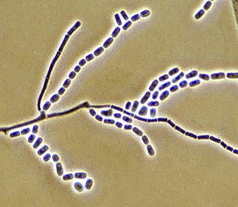
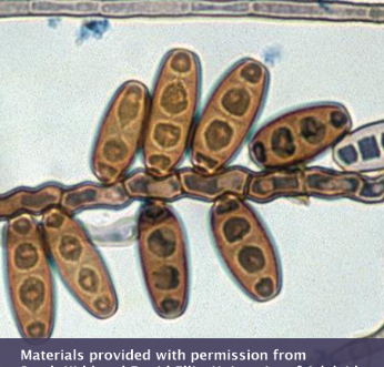
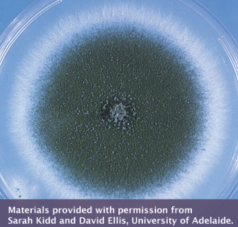
Laboratory Diagnosis of Pythiosis
ICMR Issues C. auris Advisory
Strengths and Limitations of Imaging for Diagnosis of IFI
Candidemia: Lessons Learned from Asian Studies for Intervention
Spotting invasive pulmonary aspergillosis in COVID-19 patients
Pivotal Asian Invasive Mold Study
Impact of the COVID-19 pandemic on IFI epidemiology and trends
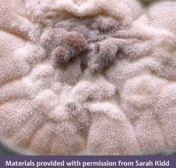
Mycetoma in Asia: Still veiled in mystery
Cryptococcosis
Identifying IFI risk factors in patients with COVID-19
ASID ANZMIG x AFWG: Fungal Frontiers in the Asia Pacific – Webinar 2
New Antifungal Agents
Gilead IFI Masterclass: Current updates on the management of IFIs in immunocompromised hosts
The AFWG Masterclass: Advanced fungal education at your fingertips
A challenging case: A crisis unfolds
The role of antifungal stewardship in improving IFI outcomes
Making Precise Diagnoses: Experience from the Laboratory Skills Enhancement Course
A challenging case: A 68-year-old man with nasal and palatal ulcers
AFWG Online Education Module 3: Optimizing Dosing in IFI Management
AFWG Online Education Module 2: Antifungal Prophylaxis in Solid Organ Transplantation
AFWG Education Module 1: The Value of Clinical Mycology Laboratories
How do I interpret Candida in the urine?
How do I interpret Candida in respiratory tract cultures?
Cryptococcosis in HIV and non-HIV infected patients
Human Pythiosis
AFWGOnline Privacy Policy has been Updated
Management of fungal infections in high-risk patients
Striving for Perfection: Experience from the Laboratory Foundation Training Course
Know your fungal landscape in Vietnam
Recent Advances of Fungal Diagnostics in Asian Laboratories
Deep Dermatophytosis: A Case Report
Management of cryptococcosis and talaromycosis
A challenging case: A 49-year-old woman with sarcoidosis
Emerging yeast infections in Asia
Outbreak of Superbug Candida auris: Asian Scenario and Interventions
Championing Medical Mycology: Thoughts on the AFWG Laboratory Skills Enhancement Course
Mucormycosis and Pythiosis – New Insights
AML and the high risk of multiple infectious complications
Do We Need Modification of Recent IDSA & ECIL Guidelines while Managing Patients in Asia?
A hospital’s experience with candidemia and empirical therapy
Top 5 most viewed AFWG videos on YouTube
Fungal Academy 2015
Fluconazole in 2015
Fungal isolation protocol
Influencing Aspergillus
Fungal Asthma
Aspergillus
Laboratory Diagnosis of IPA
Two-Hot-to-Handle
Voriconazole
Educational Organizations
Literature Updates
 

Past Quiz Results


Share this
page


Share
this page

This Exophiala species is a recognized causative agent of mycetoma and phaeohyphomycosis worldwide. In the photo, this Exophiala complex shows annellides, conidia and conidiogenous pegs (annellides) on torulose hyphae. What is this complex?

1)Exophiala jeanselmei complex
2)Exophiala dermatitidis complex
3)Exophiala spinifera complex

Exophiala jeanselmei has a worldwide distribution and is a recognized causative agent of mycetoma and phaeohyphomycosis in humans. Conidiogenous cells are predominantly annellidic and erect, multicellular conidiophores are absent. There is no growth at 40°C.

The macroconidia of this Fusarium complex are fusiform, slightly curved, pointed at the tip, mostly 3 septate, basal cells pedicellate, measuring 23–54 μm x 3–4.5 μm. This complex accounts for about 20% of human fusarium infections. What Fusarium species complex is this?

1)Fusarium fujikuroi complex
2)Fusarium chlamydosporum complex
3)Fusarium oxysporum complex

The Fusarium oxysporum complex contains at least 5 phylogenetically distinct species and accounts for about 20% of human infections caused by fusaria. It can cause localized or deeply invasive life-threatening infections in humans and other animals.

The majority of Scedosporium infections are mycetomas, but can also involve the eyes, ears, CNS and lungs. Which Scedosporium species is in the photo?

1)Scedosporium apiospermum
2)Scedosporium boydii
3)Scedosporium aurantiacum

Scedosporium apiospermum and Scedosporium boydii (formerly Pseudallescheria boydii) are now recognized as separate species and along with S. aurantiacum are the principal human pathogens. Colonies of S. apiospermum are fast growing, greyish-white, suede-like to downy with a greyish-black reverse. Unless confirmed by DNA sequencing, reporting as ‘Scedosporium apiospermum complex’ is recommended.

This Giemsa-stained touch smear shows the typical septate yeast-like cells (arrow) of which fungus? This species exhibits thermal dimorphism, is endemic in Southeast Asia and southern China, and its infection is characterized by subcutaneous abscesses and papule-like ulcers.

1)Penicillium chrysogenum
2)Talaromyces marneffei
3)Talaromyces proteolyticus

Previously known as Penicillium marneffei, T. marneffei exhibits thermal dimorphism and is endemic in Southeast Asia and southern China. Talaromyces marneffei is the only dimorphic species of Talaromyces (or Penicillium), which grows as a yeast at 37°C. It produces a red soluble pigment on general media and conidiophores have flask-shaped to acerose phialides.

Which species of Mucor (in photo) differs from other species in its formation of short circinated, branched sporangiophores bearing brown sporangia and its ability to assimilate ethanol and nitrates?

1)Mucor ramosissimus
2)Mucor amphibiorum
3)Mucor circinelloides

The genus Mucor has about 50 taxa, but only a few thermotolerant species are of medical importance. Most infections reported list M. circinelloides and similar species such as M. indicus, M. ramosissimus, M. irregularis and M. amphibiorum as pathogens.

Rhizopus arrhizus (pictured here) is one of 2 species that are the most common causative agents of mucormycosis. What is a common (but now obsolete) synonym for R. arrhizus? 1) Rhizopus oryzae 2) Rhizopus microsporus 3) Rhizopus azygosporus

1)Rhizopus oryzae
2)Rhizopus microsporus
3)Rhizopus azygosporus

The controversy surrounding which species name to use for Rhizopus oryzae versus Rhizopus arrhizus has been resolved in favor of the latter. The important medical pathogens are reduced to just R. arrhizus and R. microsporus, which account for ~60% of the reported mucormycosis cases.

This is a typical pyriform-shaped sporangium with a conical-shaped columella and pronounced apophysis (arrow) of which opportunistic pathogen? It’s known to cause pulmonary, central nervous system and skin infections in animals and humans with impaired immunity.

1)Apophysomyces elegans
2)Mucor circinelloides
3)Lichtheimia corymbifera

The genus Lichtheimia currently contains 5 mostly saprophytic plant decaying and soil-borne species. Lichtheimia corymbifera is the principle pathogen causing human and animal infections. Colonies are fast growing, floccose, white at first becoming pale grey, and up to 1.5 cm high.

These pink to dark brown colonies have been linked to cases of subcutaneous infections, keratitis, sinusitis, peritonitis and canine osteomyelitis. What fungus is this?

1)Coniochaeta hoffmannii
2)Phialemonium obovatum
3)Acremonium recifei

Colonies of Coniochaeta hoffmannii are flat, smooth, moist, pink to orange, with regular and sharp margin; reverse pink. It also forms adelophialides like Phialemonium, but in Coniochaeta, these conidiogenous cells show conspicuous collarettes, and the colonies are usually pink-salmon to dark brown.

The fungus in this photo, showing rectangular arthroconidia separated by disjunctor cells, is typically found in California’s San Joaquin Valley region and Mexico, and can cause ‘valley fever’. What is it?

1)Gymnoascus reesii
2)Coccidioides immitis
3)Paracoccidioides brasiliensis

Coccidioides immitis is typically geographically limited to California’s San Joaquin Valley region and Mexico. Microscopy shows single-celled, hyaline, rectangular to barrel-shaped, alternate arthroconidia, 2.5–4 x 3–6 μm, separated from each other by a disjunctor cell. This fungus now appears to be expanding its range within the USA and has been found as far north as the Washington State.

Brought to you by Sarah Kidd and David Ellis

The key microscopic feature of Pleurostomophora richardsiae (formerly Phialophora richardsiae) is production of:

1)Two conidial types
2)Two types of phialides
3)Both 1) and 2)

P. richardsiae is characterized microscopically by (1) hyaline conidia that are allantoid or cylindrical, and form on inconspicuous, peg-like phialides on thin-walled hyphae; and (2) brown, thick-walled conidia that are spherical to subspherical and form on dark brown, slender, tapering phialides with flaring collarettes.

Brought to you by Sarah Kidd and David Ellis

These images show the colony appearance and microscopic morphology of a common environmental mold that is emerging as a causative agent of hyalohyphomycosis in immunocompromised patients. It is:

1)Aspergillus flavus
2)Penicillium verrucosum
3)Paecilomyces variotii

Key features of Paecilomyces are long, slender, divergent phialides and culture pigmentation. P. variotii colonies are powdery to suede-like, funiculose or tufted, and yellow-brown or sand-colored. Microscopic examination will show conidiophores bearing dense, verticillately arranged branches bearing phialides.

Brought to you by Sarah Kidd and David Ellis

Exserohilum is a dematiaceous fungal genus, somewhat similar in morphology to the Bipolaris and Drechslera genera. The two photomicrographs (labeled A and B) show Exserohilum rostratum and Bipolaris australiensis. Exserohilum rostratum is shown in image:

1)A
2)B
3)Neither image

Exserohilum rostratum conidia have prominent, dark basal and distal septa, and a strongly protruding truncate hilum. Mature conidia are straight, slightly curved or bent and cylindrical to rostrate. They typically contain 7 to 9 pseudosepta (range, 4 to 14).

Brought to you by Sarah Kidd and David Ellis

This species is the only known dimorphic species in the genus Talaromyces. It produces filamentous growth at 25°C and a yeast phase at 37°C. It is:

1)T. helicus
2)T. byssochlamydoides
3)T. marneffei

T. marneffei (formerly Penicillium marneffei) is endemic in Southeast Asia. It produces a distinctive red soluble pigment on general media and exhibits thermal dimorphism. On brain heart infusion agar containing blood, incubated at 37°C, colonies are rough, glabrous, tan-colored and yeast-like.

Brought to you by Sarah Kidd and David Ellis

Histoplasma capsulatum exhibits thermal dimorphism taking on distinct forms at different incubation temperatures. This photomicrograph shows the organism growing:

1)At 37°C as a budding yeast-like fungus
2)At 25°C in its mycelial form
3)None of the above

This shows the microscopic morphology of the saprophytic or mycelial form of H. capsulatum with its characteristic large, rounded, single-celled, tuberculate macroconidia and smaller microconidia. As H. capsulatum cultures are a severe biohazard, either exoantigen test or DNA sequencing is preferred for lab safety.

Brought to you by Sarah Kidd and David Ellis

Most of the identified human opportunistic Fusarium pathogens belong to the F. solani complex, F. oxysporum complex and F. fujikuroi complex. This image shows a colony of:

1)F. solani complex
2)F. oxysporum complex
3)F. fujikuroi complex

F. oxysporum complex colonies have white aerial mycelium, becoming purple, with discrete orange sporodochia present in some strains; reverse is hyaline to dark blue or dark purple.

Brought to you by Sarah Kidd and David Ellis

This is a colony of Exophiala spinifera complex. The best medium to grow this organism is:

1)Blood agar at 35.0°C
2)Potato dextrose agar at 26°C
3)Sabouraud glucose agar at 42°C

The fungi belonging to Exophiala and related genera are still not clearly defined, and remain difficult to identify. Because of their phenotypic plasticity, it’s essential to examine isolates on media like potato dextrose agar to promote the formation of hyphal elements, and to use slide culture preparations.

Brought to you by Sarah Kidd and David Ellis

This image shows a colony of Phaeoacremonium parasiticum. The maximum growth temperature of P. parasiticum is:

1)30°C
2)40°C
3)47°C

P. parasiticum cultures are usually slow growing, suede-like with radial furrows, and initially whitish-grey becoming olivaceous-grey with age. Maximum growth temperature is 40°C. Human infections caused by P. parasiticum include subcutaneous abscesses, thorn-induced arthritis, endocarditis and mycotic keratitis.

Brought to you by Sarah Kidd and David Ellis

This is a photomicrograph of Geotrichum candidum. Which of the following is not present in the field of view?

1)Hyphae
2)Chains of arthroconidia
3)Blastoconidia

Geotrichum species do not produce blastoconidia, conidiophores or pseudohyphae. Arthroconidia of G. candidum are formed as hyphal elements are progressively compartmentalized by fragmentation of septa. Conidial secession is by schizolysis of a double septum and concomitant rupture of the original outer hyphal wall layer.

Brought to you by Sarah Kidd and David Ellis

Which of the following features is not seen in this microscopy image of Bipolaris australiensis?

1)Darkly pigmented, multicellular poroconidia
2)Long, sparsely branched chains of conidia
3)Geniculate or zig-zag rachis

This microscopy image of Bipolaris australiensis illustrates its typical features of sympodial development of pale brown, fusiform to ellipsoidal, pseudoseptate poroconidia on a geniculate or zig-zag rachis.

Brought to you by Sarah Kidd and David Ellis

Morphology-based identification methods are typically used to differentiate Aspergillus species. This Aspergillus culture is:

1)Aspergillus flavus
2)Aspergillus terreus
3)Aspergillus fumigatus Fresenius

Aspergillus fumigatus is a distinctive species that can be recognized by its broad, velutinous, bluish colonies bearing characteristic, well-defined columns of conidia.

Brought to you by Sarah Kidd and David Ellis